

Diffusion Tensor Imaging
Integral in the Diagnosis of Concussions and Traumatic Brain Injury
The Advanced Diagnostics Healthcare System (ADHS) is led by physicians who combine state-of-the-art medicine with patient-centered care. ADHS hospitals selected dedicated Texas Program- the only one of its kind in the Southwest region. Within the program, the Texas Brain Center provides best-in-class care for traumatic brain injury and brain disease. Leading-edge diagnostic technologies and procedures and the best care are available through the services at our DTI capabilities.
This technology is part of our robust Neuroscience Program, which includes Stereotactic neurosurgery (MRI), quality assurance, DTI provides quantitative assessment and diagnosis of Traumatic Brain Injury (TBI) and Cancer. For patients whose life has been impacted with brain injury and other diseases requiring symptoms, this is better—-and life-changing.
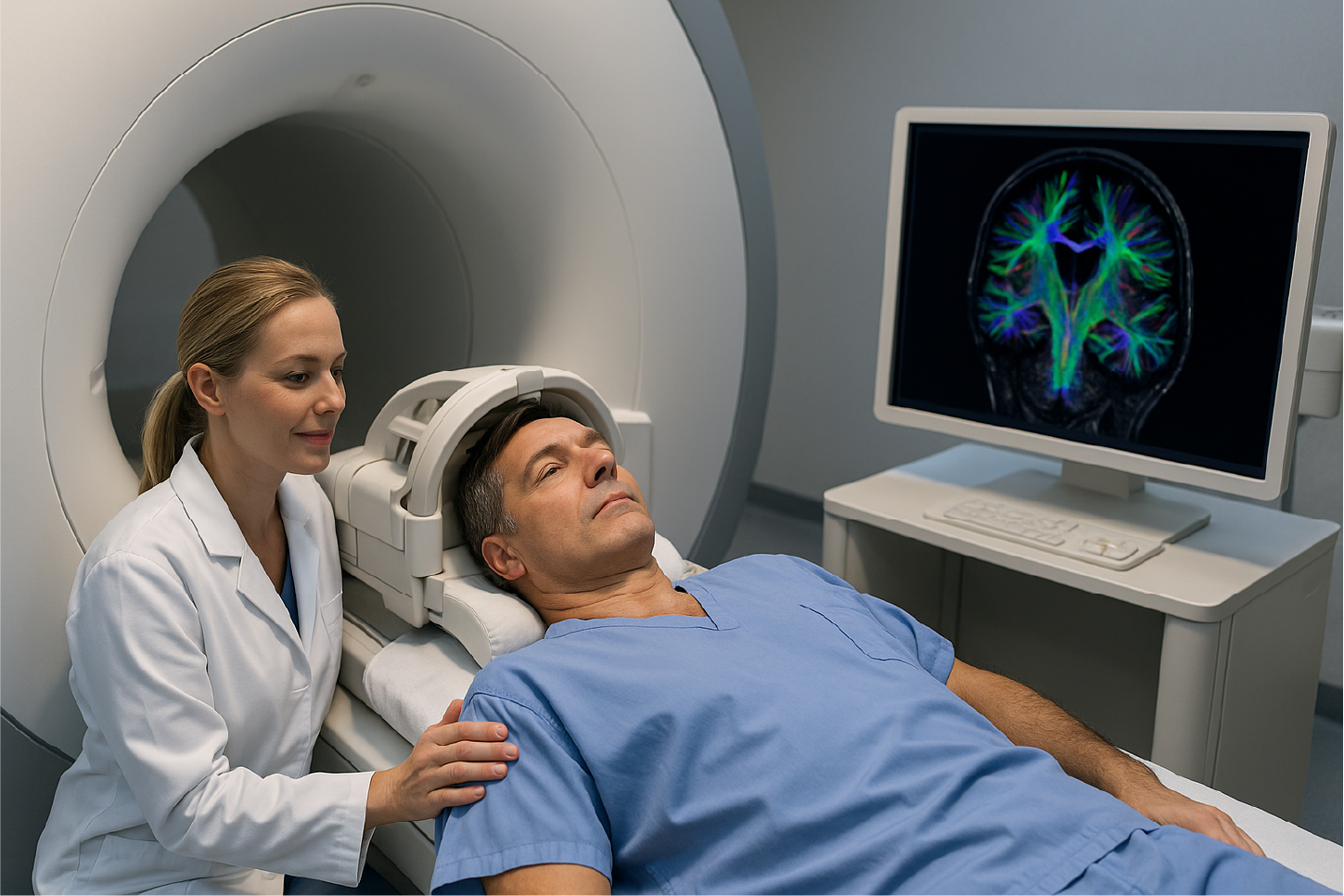
Diffusion Tensor Imaging (DTI) is a state-of-the art diagnostic tool which uses advanced MRI technology to detect traumatic brain injury that may not be visible on conventional MRI. DTI can detect damages to the brain's white matter tracts even after mild trauma, and is supplemented by brain dysfunction. DTI allows for the visualization of the brain's pathologic features while providing objective evidence of cognitive decline.
Imaging of Traumatic Brain Injury
Traumatic brain injury (TBI) is a common yet underdiagnosed problem that affects more than 1.7 million people and results in over 52,000 deaths annually. Traumatic brain injury is the most common cause of motor vehicle accidents, falls, assaults and sports-related injuries. The leading cause of death and disability, with over 85% of TBIs classified as mild, many patients may have a persisting postoncussive syndrome with 15% impairment and 20% are unable to return to work. Neuroimaging plays a critical role in the acute setting of traumatic brain injury by detecting injuries that require acute intervention such as subdural hematomas. However, in the setting of concussions and mild traumatic brain injury, conventional imaging is typically normal.
Fundamentals of DTI
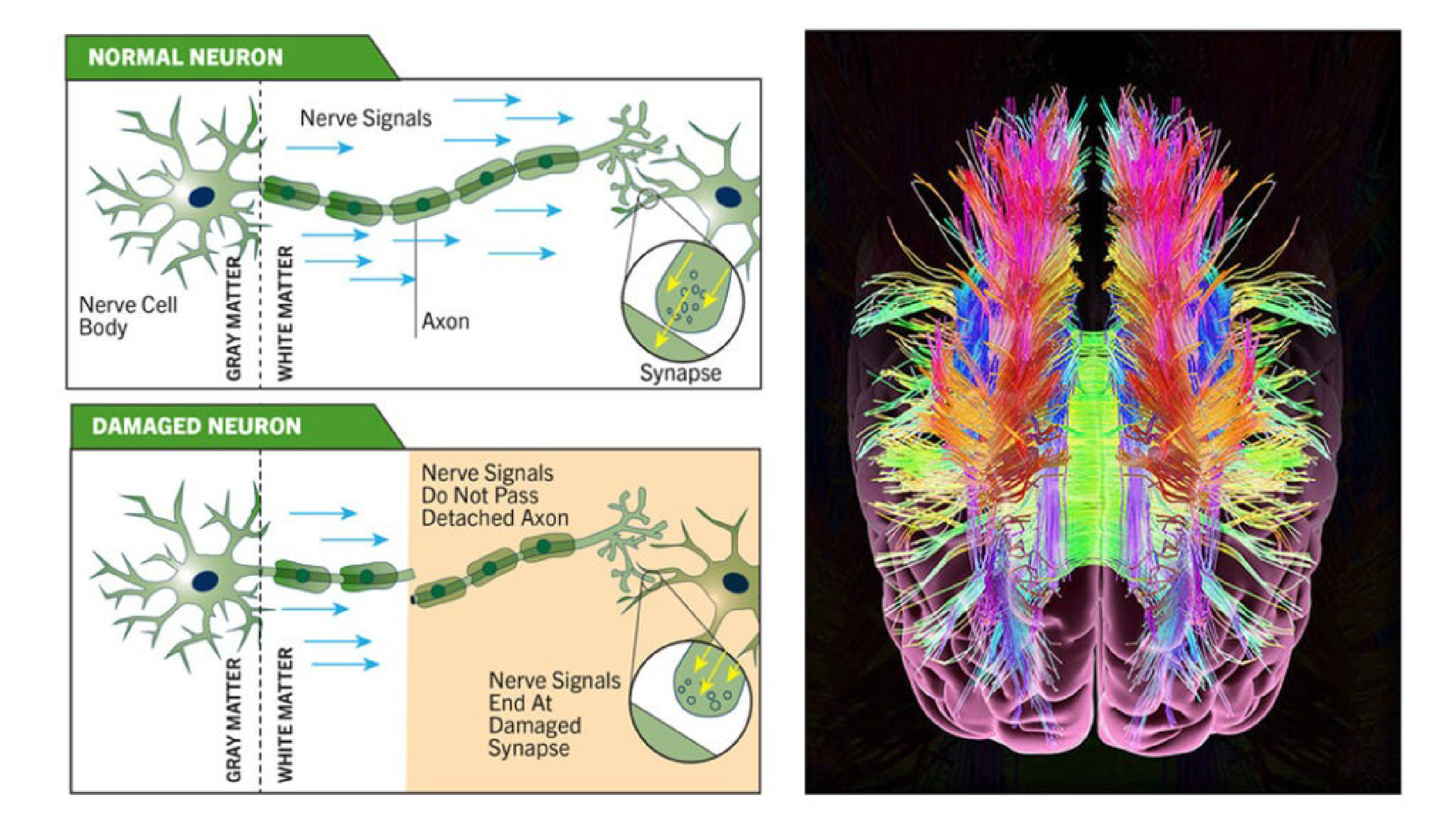
The white matter tracts of the brain are made up of neuronal fibers. They create neural pathways and bundles of thousands of axons located in either white matter areas of the brain or spinal cord (see diagram above). Axons transfer neural impulses between white matter areas. DTI measures how water moves through different brain structures.
With appropriately applied magnetic field gradients MR images can be sensitized to diffusion, or the random motion of water molecules in the tissue. Water molecules essentially random in the brain gray matter, but diffusion is anisotropic or directional in the white matter tracts. This anisotropic diffusion is because white matter axons present barriers to water motion in the directions other than parallel to the fiber tracts.
DTI tracks the degree to which the water molecules are moving with random Brownian motion. A molecule that is constrained by a white matter tract will move more in one direction, allowing structures and injury to be visualized.
In summary, detection of microstructural damage by using DTI is a major advancement in the diagnosis of TBI. Executive function, which is largely dependent on the periventricular periventricular cortex, is commonly impaired after mild TBI and is a major contributor to disability. DTI provides objective evidence of brain injury related to impairment even in the setting of otherwise normal imaging.
Available at the ADHS Hospitals.
For More Information, Contact:
Kevin Smoot - Executive Director, Subacute Trauma Program
ksmoot@SubacuteTrauma.com
713-848-4702